New X-ray Technologies and Future Advances
"Revealing the unknown. Combining neutron and X-ray imaging a turning point in matter probing"

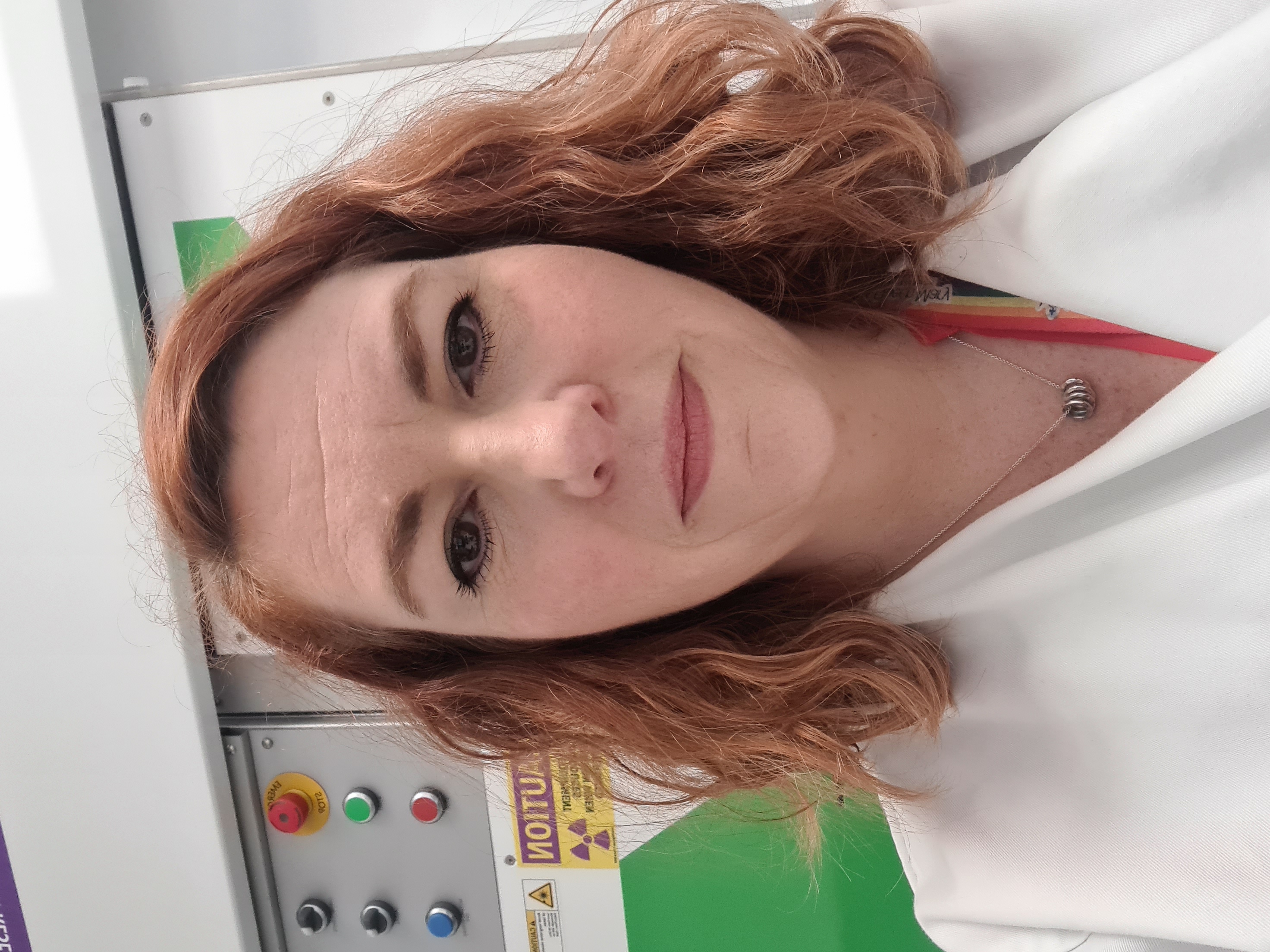
Dr Genoveva Burca, ISIS Facility, STFC, UK
Genoveva is an international expert in neutron imaging and diffraction techniques based at the ISIS Neutron and Muon Source, Oxfordshire, UK and visiting academic at the University of Manchester. Her research focuses on solving complex problems at the intersection of complementary neutron and x-ray ...
Genoveva is an international expert in neutron imaging and diffraction techniques based at the ISIS Neutron and Muon Source, Oxfordshire, UK and visiting academic at the University of Manchester. Her research focuses on solving complex problems at the intersection of complementary neutron and x-ray imaging techniques building robust solutions for materials characterization from a cross-disciplinary scientific perspective. Apart from her creative and interdisciplinary approach on delivering solutions to academia and industry via combined imaging techniques she also provides training and mentors the next generation of scientists in the efficient use of large scale scientific neutron facilities. She actively serves on several national and international advisory and reviewing committees and collaborates with various research groups from UK, Sweden, Denmark, Switzerland, France, Canada and India as well as industrial partners on various projects spanning from life sciences to engineering.

Life Sciences
"Towards post-mortem imaging of the entire human brain with true cellular resolution"

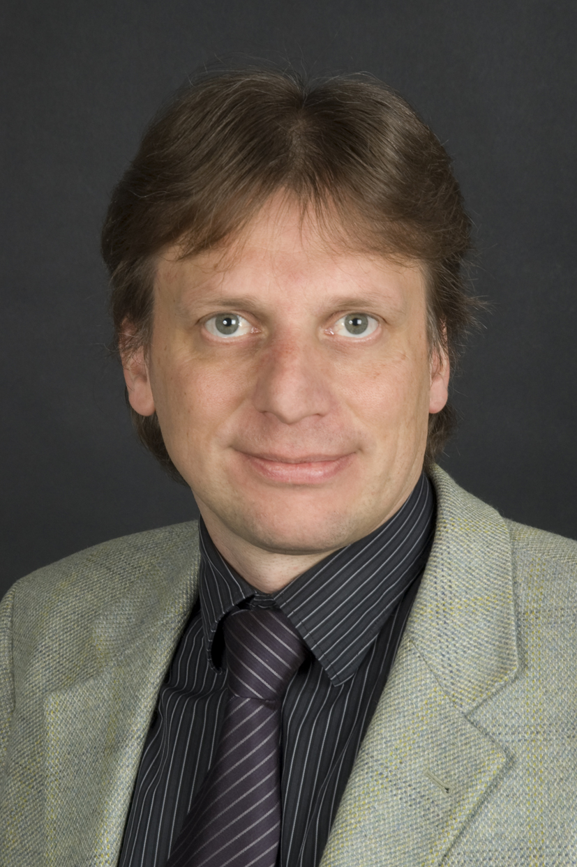
Dr Bert Müller, Universität Basel, Switzerland
In 2006, Bert Müller has been appointed as the first Thomas Straumann Chair for Materials Science in Medicine at the University of Basel, Switzerland. He is the founding director of the Biomaterials Science Center – an integral part of the oldest medical faculty in Switzerland. Since 2001 he has ...
In 2006, Bert Müller has been appointed as the first Thomas Straumann Chair for Materials Science in Medicine at the University of Basel, Switzerland. He is the founding director of the Biomaterials Science Center – an integral part of the oldest medical faculty in Switzerland. Since 2001 he has been independently teaching at the physics department of ETH Zurich. In addition, Bert Müller has been one of the maximal one hundred research councillors of the Swiss National Science Foundation since 2019. In 1989, he received his Master degree in physics from the Dresden University of Technology, Germany. He earned a doctoral degree in experimental physics from the University of Hannover, Germany, in 1994. The research output is characterized by almost 400 journal papers and book chapters. Bert Müller was the main supervisor of 35 doctoral thesis projects. His current research interests include high-resolution hard X-ray imaging and physics-based approaches to challenges in medicine and dentistry. For the fourth time, he is the chair of the well-established conference “Developments in X-ray Tomography”. Bert Müller has been named the 2022 recipient of the SPIE Biophotonics Technology Innovator Award in recognition of his distinguished contributions to 2D and 3D imaging for the challenges of the 21st century including cardiovascular and neurodegenerative diseases. Most recently, he has developed to an entrepreneur: The spin-off companies Bottmedical, Acthera Therapeutics and Bottneuro were founded in 2019 and 2021.

Correlative Imaging
"Correlative tomography and its applications in the Preclinical Imaging Facility at Barts Cancer Institute."

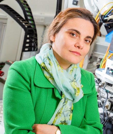
Dr Julie Foster, Queen Mary University London, UK
Julie is a biologist who completed her PhD in physiology from the University of Canterbury in New Zealand quite some time ago. Past highlights include; work on the cardiovascular physiology of the primitive and slimy hagfish, virology studies in ferrets with flu and therapeutic studies in rats with ...
Julie is a biologist who completed her PhD in physiology from the University of Canterbury in New Zealand quite some time ago. Past highlights include; work on the cardiovascular physiology of the primitive and slimy hagfish, virology studies in ferrets with flu and therapeutic studies in rats with renal failure. She currently manages the Preclinical Imaging Facility at BCI and works in the Centre for Cancer Biomarkers and Biotherapeutics at Queen Mary. Julie enjoys working with both academic and industry partners to optimise their imaging studies, no project is ever the same. With a well-appointed imaging facility able to offer a range of imaging modalities (including SPECT, PET, CT, MRI, bioluminescence, fluorescence and ultrasound), correlative tomography offers an opportunity to develop techniques and answer questions that couldn’t be addressed using a single imaging modality alone.

Cultural Heritage

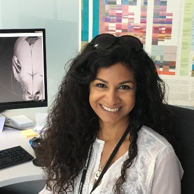
Professor Anjali Goswami, Natural History Museum, UK
I am currently Dean of Postgraduate Education and Research Leader in Life Sciences at the Natural History Museum, London, as well as Honorary Professor in Palaeobiology in the Department of Genetics, Evolution, and Environment at University College London. My main research interests are in ve...
I am currently Dean of Postgraduate Education and Research Leader in Life Sciences at the Natural History Museum, London, as well as Honorary Professor in Palaeobiology in the Department of Genetics, Evolution, and Environment at University College London. My main research interests are in vertebrate evolution and development, especially using 3D morphometric methods to incorporate data from embryos to fossils to test genetic and developmental hypotheses of modularity and morphological diversity and reconstruct macroevolutionary patterns through deep time. I have previously focused on skull evolution in carnivorans and the marsupial-placental dichotomy, but in recent years, I have expanded across the diversity of systems, with an ERC-funded project on modularity and morphological diversification across tetrapods, a Leverhulme Trust-funded project on pelagic fish skull evolution, a recently-completed Leverhulme Trust-funded project on cat postcranial mechanics and evolutionary modularity, and multiple projects focused on whale skull evolution and development and multivariate methods development. With collaborators and lab members, we are currently extending this work to incorporate comparative genomic and transcriptomic data, as well as applying our morphometric innovations to comparative ontogenetic morphology. I am also currently working on the relationships and paleobiogeography of Mesozoic and early Cenozoic mammals, particularly focusing on Gondwanan eutherians, with a recently-completed Leverhulme Trust-funded project uniting genomics and fossils to elucidate early placental evolution. As part of this work, I was also involved in a Leverhulme Trust-funded project on the evolution of latitudinal biodiversity gradients in Cretaceous vertebrates. I currently conduct fieldwork in the Cretaceous and Palaeogene of India and Argentina, but have previously been involved in fieldwork in Svalbard, Peru, Chile, Madagascar, and the United States. I have also conducted palaeoecological research on Triassic amniotes and Eocene whales using dental microwear and stable isotopes.

Life Sciences
“Micro-CT-based 3D virtual histology for forensic, medical and veterinary entomology research”

Dr Daniel Martín-Vega, Universidad de Alcalá, Spain
Daniel Martín-Vega is currently a Lecturer in Zoology and Entomology at the University of Alcalá (Madrid, Spain) and a researcher at the IUICP (University Institute of Research in Police Sciences). His research interests focus on the study of insects of forensic, medical and veterinary relevance. ...
Daniel Martín-Vega is currently a Lecturer in Zoology and Entomology at the University of Alcalá (Madrid, Spain) and a researcher at the IUICP (University Institute of Research in Police Sciences). His research interests focus on the study of insects of forensic, medical and veterinary relevance. Daniel started using micro-computed tomographic techniques during a two-year stay at the Natural History Museum (London) with the support of an EC-funded Marie Skłodowska-Curie fellowship, in order to develop a novel method for aging blow fly puparia to estimate the time of death in forensic casework. Since then, he has also applied micro-CT to the study of parasite/host interfaces in medical and veterinary entomology research. Daniel is author or co-author of more than 50 peer-reviewed publications and he has participated in 10 research projects funded through competitive calls. Daniel is a Scientific Associate at the Natural History Museum, a Fellow of the Royal Entomological Society, and a member of the AeE (Spanish Association for Entomology) and EAFE (European Association for Forensic Entomology).

Buy Tickets

Submitted an abstract? Don’t forget to register your attendance!
Attend conference